


3D Printed Metastatic Adenocarcinoma in the Brain
Clinical History
A 56-year old male underwent a total gastrectomy and splenectomy for gastric
adenocarcinoma. Over a period of two months he developed a progressively unsteady gait,
increasing weakness of his left hand and frontal headaches associated with nausea and vomiting.
Imaging revealed a lesion in the right frontal lobe. He underwent a craniotomy with resection of
the lesion, which was confirmed metastatic gastric adenocarcinoma. He experienced gradual
increasing symptoms as well as jaundice, deteriorating consciousness and papilloedema from
increased intracranial pressure. Repeat imaging revealed recurrence of the right frontal
metastatic lesion as well as liver metastases. The patient died 9 months after his initial
gastrectomy surgery.
Pathology
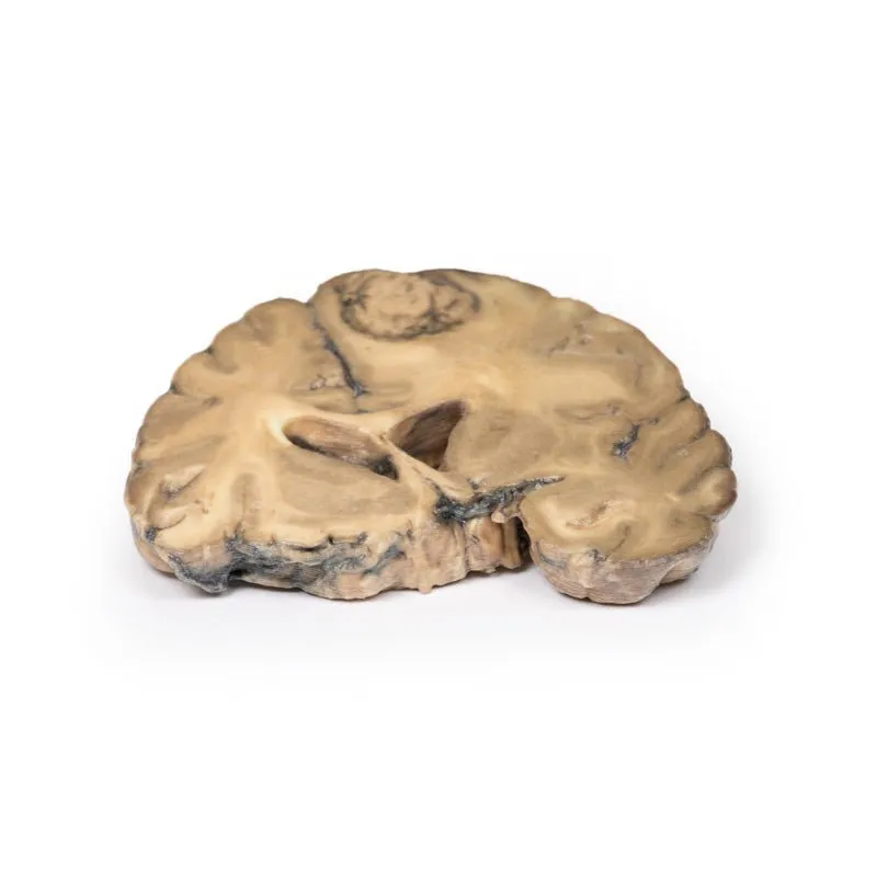
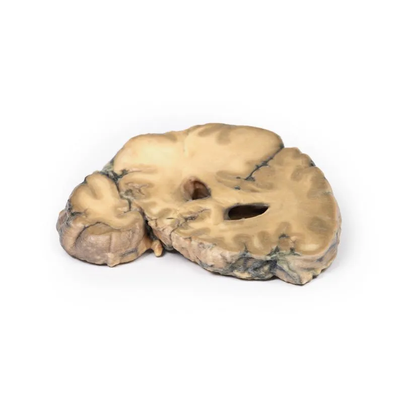
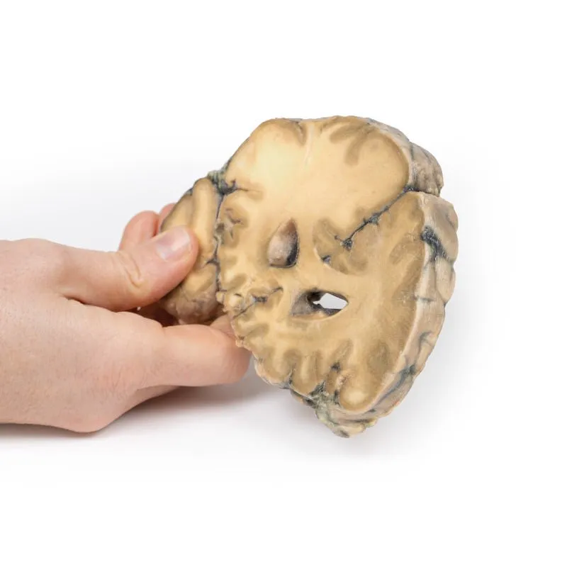
This brain specimen is cut in the coronal plane. A circumscribed, variegated,
pink-grey tumour is evident in the right frontal lobe. The tumour is involving the grey and
white matter. Compression of the right lateral ventricle by the lesion is apparent with shift of
the midline structures also seen.
Further Information
Stomach cancer is one of the most common causes of cancer-related death
worldwide. Risk factors include male gender, diet, smoking and chronic Helicobacter pylori
infection. The most common sites for metastases of gastric adenocarcinoma are the liver,
peritoneum, lungs and bones. Brain metastases are rare, occurring in <1% of cases. Isolated
brain metastases are very uncommon with them being more commonly seen in disseminated disease
and associated with a poor prognosis. Palliative treatment may include surgery, radiotherapy,
steroid, chemotherapy or a combination thereof.
GTSimulators by Global Technologies
Erler Zimmer Authorized Dealer


 - Mint")


